In September 2026, Google Research, HHMI Janelia, the University of Cambridge, and the MRC Laboratory of Molecular Biology published the largest brain map by neuron count to date: the complete connectome of the adult male fruit fly's central nervous system.
It contains 166,000 neurons and 125 million synaptic contact points, resolving into roughly 25 million distinct neuron-to-neuron connections. The map covers the central brain, the optic lobes, and the ventral nerve cord, the fly's equivalent of a spinal cord. The project began in 2008 and took nearly two decades to finish. Electron microscopes sliced the brain into thousands of ultra-thin sections, and AI-assisted software stitched the images back into a single wiring diagram. The goal was medical: understand how healthy circuits process behaviour, learn how damaged ones might be repaired, then move to a mouse brain and eventually a human one.
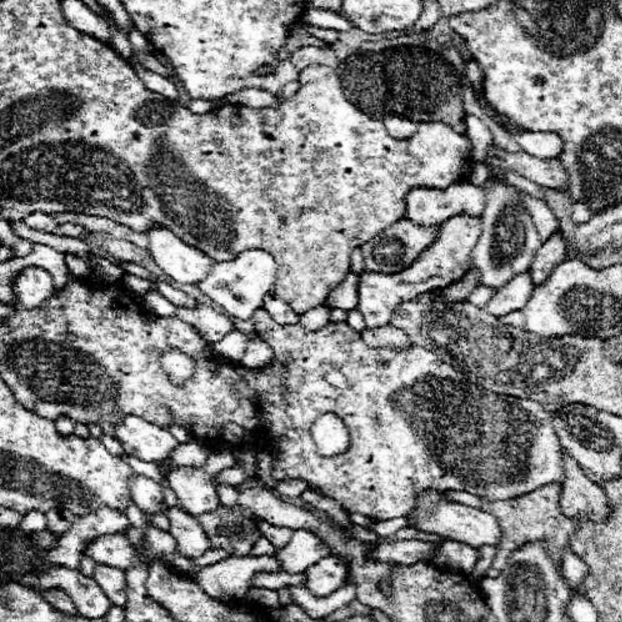
One 8-nanometre electron micrograph section through the adult male fly's brain, a single slice from the volume the connectome was traced out of. Image: EM data from the MaleCNS v1.0 dataset, FlyEM (HHMI Janelia), University of Cambridge, MRC Laboratory of Molecular Biology and Google Research. CC BY 4.0, cropped.
Three days later, it was playing Doom
It was published under an open licence and structured as a network graph. Three days after the paper, a hobbyist project called doomfly had the male fly's connectome driving a character through Doom. Within the week, others had it playing Super Mario 64 and Minecraft. One demo wired the connectome into an email classifier and reported roughly 80% accuracy on that task, while conceding it still trailed a small conventional neural network. Across fly-connectome demos generally, results have ranged from close to chance to above 90%.
None of this was the point. The connectome was built to study pathway repair, not to run a platformer. But data released under an open licence behaves like any public dataset: people use it for what it can technically do, not only for what it was designed to do. Decades of medical research became a novelty in days.
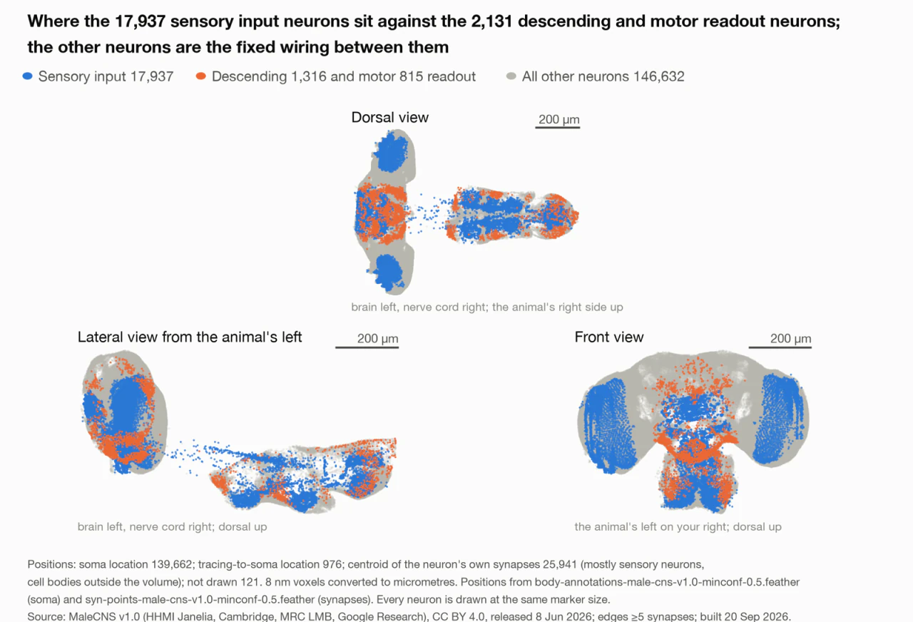
All 166,700 classified neurons of the male fly's central nervous system and ventral nerve cord, plotted in three views and coloured by functional class. This is the dataset the hobbyist demos loaded. Image: Jason Alan Snyder, SuperTruth; data: MaleCNS v1.0 (HHMI Janelia, Cambridge, MRC LMB, Google Research). CC BY 4.0.
A connectome is a graph, not a recording
A connectome is a complete wiring diagram: every neuron and every connection between them, mapped in full. It records what is connected to what. It does not record what the brain is thinking.
Three regions make up the fly's map. The central brain handles core processing. The optic lobes process visual input from the compound eyes. The ventral nerve cord relays signals between brain and body. Every neuron falls into one of two functional roles: sensory neurons carry information in, motor neurons carry commands out. Those roles matter later, because they are the only points where an outside system can attach to the fly's circuitry.
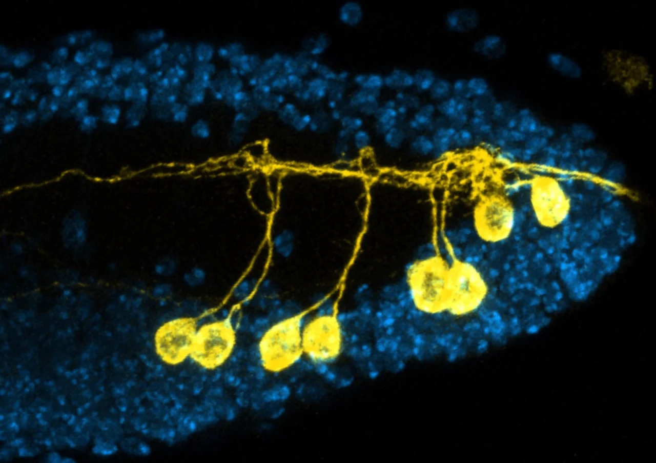
Neurons of the fly's ventral nerve cord under a confocal microscope, glowing against a field of cell nuclei. The ventral nerve cord relays signals between brain and body, much as a spinal cord does. Image: Gerit Linneweber, University of Cambridge. CC BY 2.0, via Wikimedia Commons.
Structurally, the dataset is a graph. Neurons are nodes; synapses are edges. That is the representation computer scientists already use for networks of any kind, which is why a biological wiring diagram loads into standard graph software with almost no translation.
Loading the graph solves half the problem. A fly's eyes never saw a screen, and its legs never touched a controller. Something has to translate a game's pixels into a signal the fly's sensory neurons can accept (encoding), and translate the resulting motor activity back into an action such as "move forward" (readout). Both translations are human decisions. There is no scientifically correct way to encode a Minecraft screen into fly-eye input; whoever builds the demo chooses. The wiring is real. The behaviour it produces in a game is an interpretation layered on top by a person.
That distinction makes the label harder to apply. If no person designed these connections to solve a task, calling the result artificial intelligence needs more than a resemblance.
These weights were never trained
In machine learning, a weight is a number that controls how much one signal influences another as it passes through a network. Multiply the input, sum the results, and the weights determine the output. Those weights start out meaningless and are made meaningful by training: a network sees an example, compares its output with the correct answer, calculates the error, and adjusts every weight slightly to reduce it. That is backpropagation, paired with gradient descent. Repeated across millions of examples, the weights converge on something useful. A modern model can hold hundreds of billions of them.
This connectome has weights too, and they were never trained. Each is a direct physical measurement: the number of synaptic contact points between two specific neurons, counted from electron microscope images. No loss function, no gradient descent, no target output to optimise toward. The value is what evolution and one fly's biology produced.
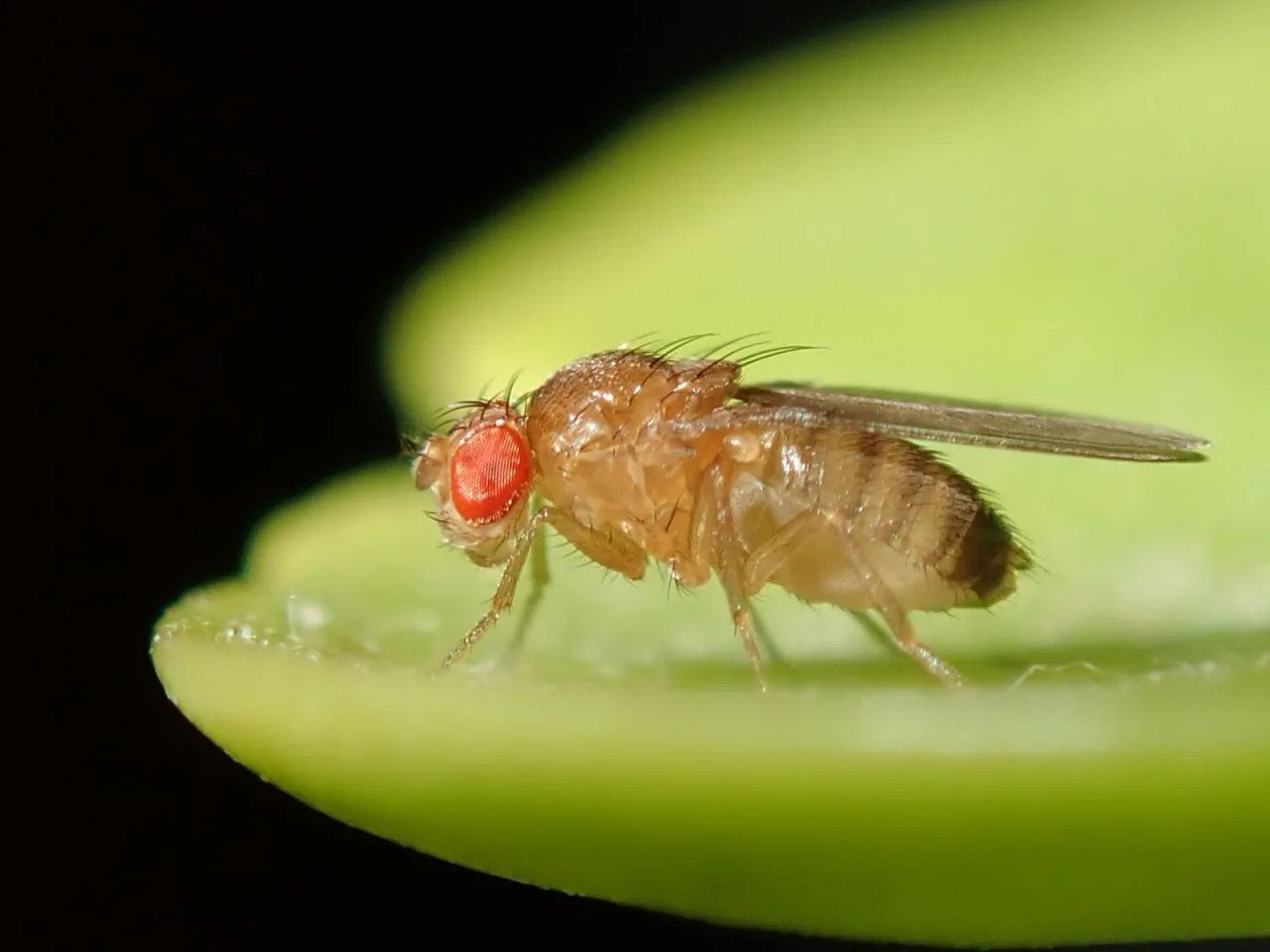
Drosophila melanogaster. Every "weight" in its connectome is a measured count, not a learned parameter. Image: Alexis (alexis_orion) via iNaturalist and Wikimedia Commons. CC BY 4.0.
That is the distinction that matters. A pretrained model and this connectome share a computational shape: nodes, weighted edges, forward propagation. One set of weights was optimised for a task; the other is a snapshot of anatomy, repurposed to behave like an optimised system. The closer comparison is a circuit board that nature pre-wired, with values read off the hardware instead of learned from data.
Those values did not come from computer science. They came from biology.
Three forces, three timescales
Three forces set the numbers in this connectome, each on a different timescale.
Evolution acts across generations. Over millions of years, natural selection shaped the basic circuit plan of the fly's nervous system: which neuron types exist, how they broadly connect. It encoded that plan in the genome.
Development acts within a single fly, largely before experience begins. Genes guide neurons to grow and form initial connections from that inherited blueprint.
Neuroplasticity acts during the fly's life. Synapses strengthen or weaken with real activity and experience, the same mechanism that underlies learning in any brain.
All three leave marks on the structure the microscope captured, and the scan cannot separate inherited blueprint from lived experience. In insects the balance skews heavily toward the first two: flies rely far less on experience-driven rewiring than mammals do. What plasticity exists concentrates in one region, the mushroom body, the fly's learning and memory centre.
This is not a generic template. It is a snapshot of one individual fly at one moment, carrying whatever mix of inheritance and personal history that fly had accumulated. Lived experience is written into synaptic strength, and those strengths were captured in full.
Did the scan also capture what the fly remembered?
Memory, recorded but unreadable
In flies, memory lives in synaptic strength. A learned association, such as an odour paired with danger, becomes a physical change to specific connections in the mushroom body. Learn something, and the relevant synapses shift.
Every synaptic strength in the fly's brain was captured at the moment it was scanned. If this fly learned anything during its life, those adjustments are in the data, indistinguishable in the raw numbers from ordinary baseline wiring.
Baked in is not the same as retrievable. Nothing labels which of the 125 million contact points reflects a memory rather than default anatomy. There is no record of what the fly experienced, which odour, or what outcome. It is a hard drive with data on it and no file format to read it back.
Recall makes this worse. A real memory requires the original cue, delivered the way the fly's biology would have received it, processed through circuitry in its original chemical state. Feed that same circuit a Minecraft screen instead, translated through an arbitrary encoding, and the odds it matches the cue the fly once learned are effectively zero. The lock may still exist. The key was never cut.
At most, a faint structural bias might linger: a diffuse influence on how signals propagate through that region. Not a memory surfacing, not recognition. Residue with no address. The physical trace may be there; the ability to summon it did not survive the scan.
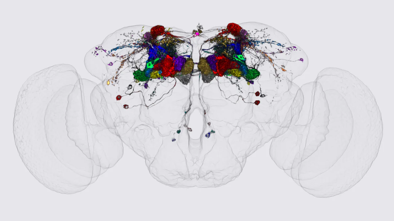
Mushroom body output neurons, segmented and shown inside a translucent fly brain. The mushroom body is where a lifetime of learned associations would have been written into the connectome. Image: eLife / Aso et al. (2014), eLife 3:e04577. CC BY 2.0, via Wikimedia Commons.
The gap to whole brain emulation
Scale the technique up and the target is the human brain. Whole brain emulation means mapping every neuron and connection of an actual mind, then running it as a simulation.
That gap is enormous. This connectome holds 166,000 neurons; a human brain holds roughly 86 billion, about 500,000 times more. Current scanning methods are also destructive: tissue is sliced into thin sections to be imaged, which rules out scanning a living person who then walks away. Whether a non-destructive method at sufficient resolution is physically possible remains unknown.
Even a perfect scan would not settle the deeper question. This is the hard problem of consciousness: structural knowledge of a brain's wiring does not establish whether the system running on that wiring experiences anything at all. No test confirms subjective experience even in principle. The connectome already demonstrated that limitation with a fly's memories: structurally present, functionally unreachable.
There is a second problem, independent of the science: personal identity. If scanning destroys the original, the person either survives as a continued mind or ends with a copy taking over. If scanning is non-destructive, two entities exist, each equally convinced of being the original. Philosophers call it the teleporter paradox, and it has no settled answer.
Whole brain emulation may well succeed as a technique: a system that behaves exactly like a specific person, given enough time and computing power. Whether that satisfies what uploading promises, continuity of self rather than a convincing impersonation, remains unresolved. The technology may arrive. Whether it delivers what anyone hoped for is a separate question, and not one engineering will answer.
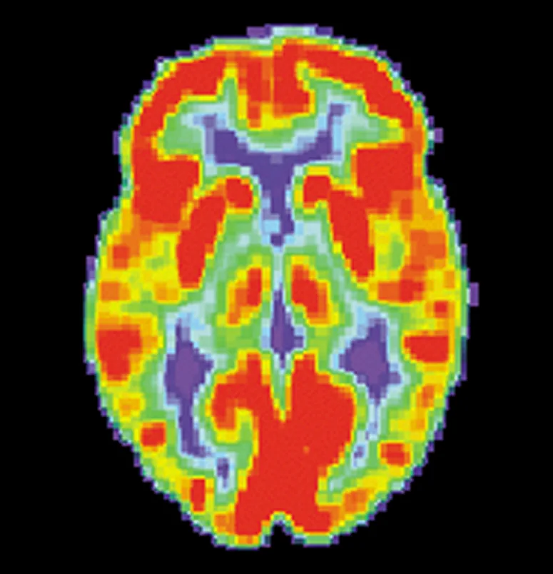
An axial PET scan of a normal human brain. Whole brain emulation would have to resolve every neuron and connection in a structure like this one, roughly 86 billion neurons. Image: Alzheimer's Disease Education and Referral Center, National Institute on Aging (US National Institutes of Health). Public domain.
Downloading it yourself
The connectome is public today at male-cns.janelia.org, under a CC-BY licence. NeuPrint provides interactive access backed by a Neo4j graph database queried through Cypher, and Python and R packages offer programmatic access with a free API token.
Format follows purpose. Researchers query the graph database directly. Developers export flat connection tables for smaller-scale analysis. Machine-learning practitioners use repackaged versions built as PyTorch-ready tensors. Each format represents the same wiring diagram, structured for a different tool.
That openness is what put a fly's brain inside Doom within days of release: a full scientific dataset, published freely, available to anyone with a connection and the curiosity to ask what else it might do. Nearly two decades of work produced a snapshot of one fly's nervous system: structure without a mind behind it, yet still capable of driving one, in a game, on an ordinary computer.